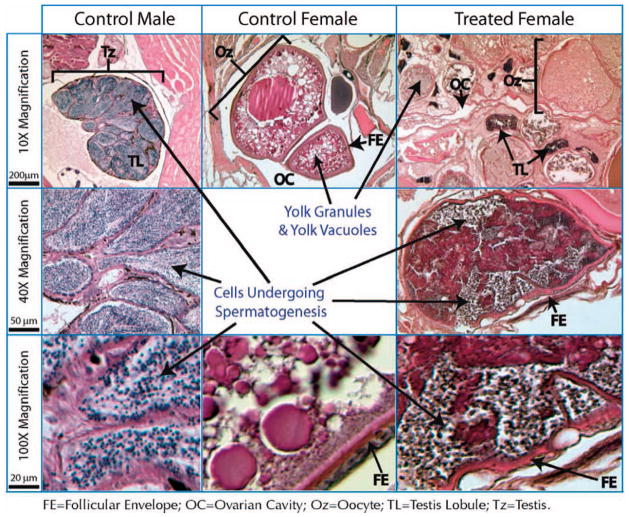
Fig. 5.
Hematoxylin and eosin–stained sections through untreated, control male and female fish along with a treated, hermaphroditic female. The left three panels show male histological features, including the entire testis (Tz), testis lobules (TL), and sperm-producing cells, stained dark blue. The center panel shows a section of a developing oocyte (Oz) in a control female, including dark red–staining yolk granules and clear yolk vacuoles. The right panels show intersexual gonads of a genotypically female fish, which appears to contain a mixture of both types of tissues, including oocytes, ovarian cavity (OC), and what appear to be sperm lobules. The higher-magnification image in the bottom right corner exhibits darkly staining sperm-producing cells, similar to what is seen in the control male testis (lower left). The histological work indicates that the intersexual gonad is a chimera of ovarial and testicular tissue.

