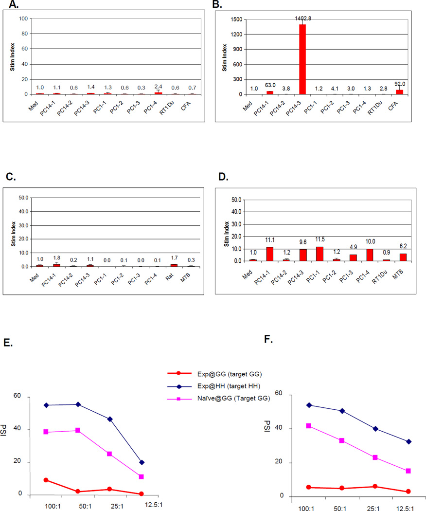
Figure 1.
Representative in vitro proliferation assays. (A) Peptide proliferation assays (PPA) to PC14 class Ic peptides in a naïve (unimmunized) animal, 16619 and (B) 21 days after this animal was immunized with each of the PC14 class Ic peptides. (C) PPA to PC1 and PC14 class Ic peptides in the long term heart and kidney recipient (#17033) before immunization and (D) after immunization with each of the PC1 and PC14 class Ic donor peptides. (E) Cell mediated lympholysis (CML) assays performed in at the same time in the same long term heart and kidney recipient (#17033) before immunization and (F) after immunization with each of the PC1 and PC14 class Ic donor peptides. In these assays, PBMCs from SLAdd (Id IId) experimental animals (DD) were either primed by irradiated SLAgg (Ic IId) stimulator cells (GG) and then incubated with chromium-labeled GG target cells or primed by irradiated SLAhh (Ia IId) stimulator cells (HH) and then incubated with chromium-labeled HH cells. As controls, PBMC’s from naïve DD animal were primed by irradiated GG cells then incubated with chromium-labeled GG target cells.

