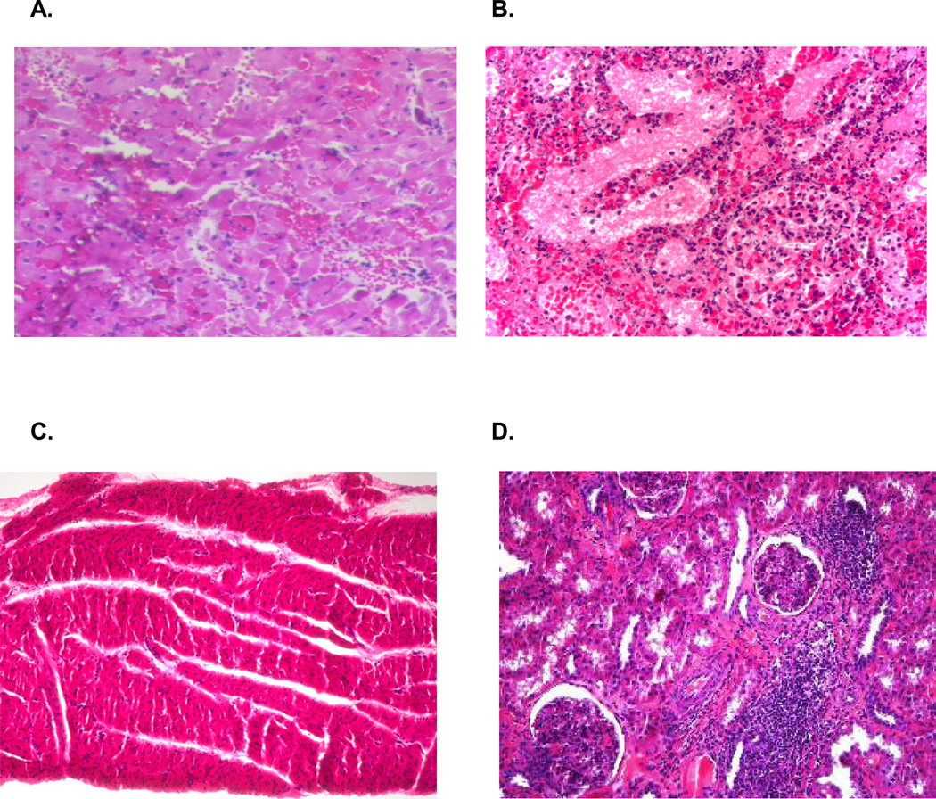
Figure 2.
Representative graft histology by hematoxylin and eosin staining. (A) Necropsy specimen from the cardiac allograft in recipient #15942 showing ISHLT grade 4/4 rejection. (B) Necropsy specimen from the renal allograft in recipient #15942 showing ACR 3 rejection. (C) Biopsy specimen from the cardiac allograft of animal #17033 biopsies on POD 196 showing no evidence of cellular rejection. (D) Biopsy specimen from the renal allograft of animal #17033 biopsies on POD 196 showing a mononuclear cell infiltrate but no signs of kidney graft destruction (ACR 1).

