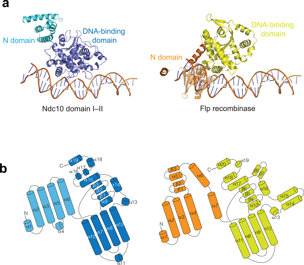
Figure 3. Structural alignment of K. lactis Ndc10 DI–II with Flp recombinases.
(a) Monomer structure of Flp (PDB ID: 1M6X) aligned with the K. lactis Ndc10 DI–II. The N-domain and the DNA binding domain of Flp recombinase are colored in orange and yellow, respectively. In Flp, the DNA structure of the Holliday junction was replaced by 30 bp CDEIII DNA for simple comparison. (b) Folding diagrams of K. lactis Ndc10 DI–II and Flp recombinase. Secondary-structure elements are labeled according to their position in the polypeptide chain; domains colored as in panel a.

