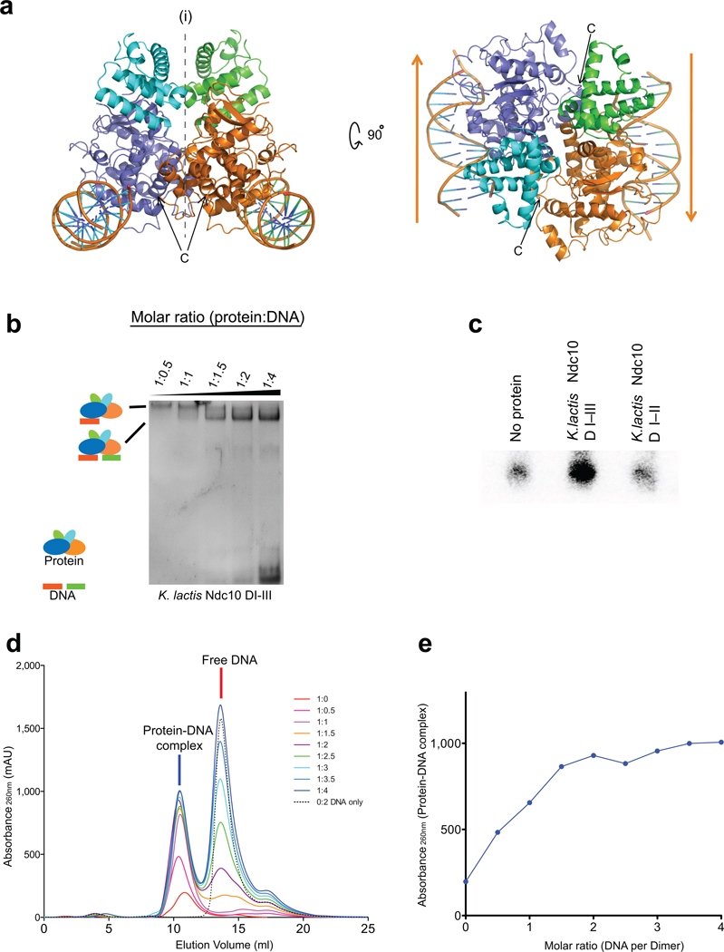
Figure 4. Dimerization of K. lactis Ndc10 DI–III.
(a) Views of the likely Ndc10 DI–II dimer (symmetry axis along b in the C2221 space group). The subunits of the dimer contact different pseudocontinuous DNA duplexes. (b) EMSA of Ndc10 DI–III with increasing amounts of 30 bp CDEIII DNA. (c) DNA capture assay with two different labels. Either Ndc10 DI-III or Ndc10 DI-II was incubated with a mixture of equal amounts of biotinylated and unmodified CDEIII DNA, the including 32P-labeled product (10%). (d–e) Ratio of Ndc10 DI–III and CDEIII DNA determined by analytical size-exclusion chromatography.

