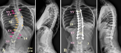
Fig. 3.
a A 14-year-old girl with a single thoracic AIS, type B, in which the NV is located at the EV + 3. The distal fusion level is the NV-1 (L2) and the direction of the DVR of the two lowest instrumented vertebrae is the same direction compared with the thoracic DVR. b The patient is treated with the rod derotation and the DVR method

