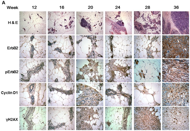
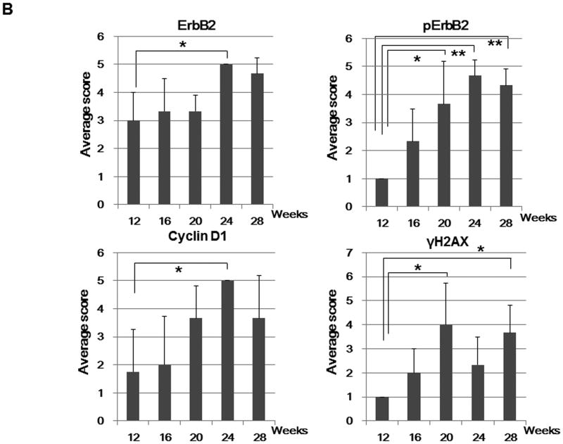
Figure 3. pERBb2 is overexpressed in BRCA-1 deficient mice.
A, Histological and immunohistochemical analysis of mammary glands and tumors over time in Brca1 Co/Co;MMTV-Cre;p53+/- mice. Paraffin sections of the mammary glands or tumors from 12, 16, 20, 24, 28, and 36 week-old Brca1Co/Co;MMTV-Cre;p53+/- mice were stained with H & E (top) and the indicated antibodies (lower, ErbB2, pErbB2, cyclin D1, and γH2AX). Original magnifications: X 100 (H & E); X 400 (immunohistochemistry). B, The percentage of cells positive for ErbB2, pErbB2, cyclin D1, and γH2AX staining was scored in a blinded manner: 1, < 5% positive cells; 2, 5-20% positive; 3, 20-50% positive; 4, 50-80% positive; 5, > 80% positive. n = 3 per group. *, p < 0.05; **, p < 0.001. C, Western blot analysis of pErbB2, ErbB2, cyclin D1, and γH2AX levels in the mammary glands from mice 12, 16 and 20 weeks of age and in mammary tumors from 35 week-old BRCA1-deficient mice.


