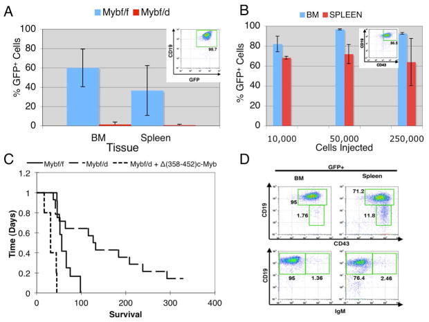
Figure 1.
c-Myb hemizygous bone marrow cells are less efficiently transformed by the p190BCR/ABL oncogene. (A) Infiltration of p190BCR/ABL-transduced Mybf/f and Mybf/d bone marrow cells into bone marrow and spleen of lethally-irradiated recipient mice. GFP expression from the MigRI/GFP bicistronic retrovirus was used to detect BCR/ABL-expressing donor cells. Recipient mice were sacrificed 8 weeks after transplantation with 2×10⁶ MigRI p190BCR/ABL-transduced bone marrow cells. GFP+ cells were detected by flow cytometry; inset shows that nearly all GFP+ cells were CD19+; (B) Different numbers of CD19+CD43+ GFP+ cells (inset) from mice transplanted with p190BCR/ABL-transduced Mybf/f marrow cells were injected into sub-lethally-irradiated secondary recipient mice and GFP-positive cells analyzed by flow cytometry in bone marrow and spleen once moribund mice were sacrificed (2–3 weeks post-injection). (C) Kaplan-Meier plot shows survival of mice injected with 2 × 10⁶ p190BCR/ABL-transduced Mybf/f or Mybf/d bone marrow cells transduced with p190BCR/ABL alone with p190BCR/ABL and Δ(358–452) c-Myb. (D) Representative immunophenotype of GFP+ cells from bone marrow and spleen of mice injected with p190BCR/ABL-transduced Mybf/f cells [CD19+CD43+ (top panel); CD19+IgM+ (bottom panel)].

