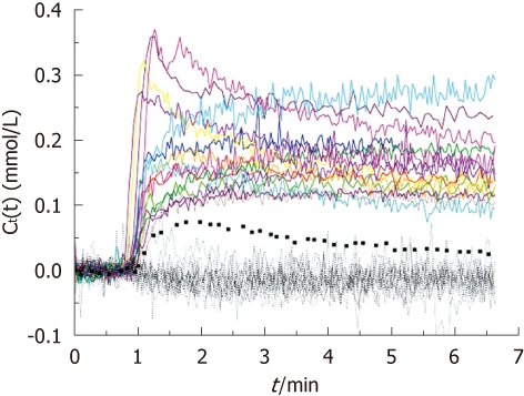
Figure 1.

Contrast agent concentration vs time curves in bone metastases (colored solid lines) and normal bones (black dotted lines). The curve from the only enhancing normal bone was highlighted by thick black dotted line. The enhancement of the normal bone was much less than the metastatic lesions.
