Figure 4.
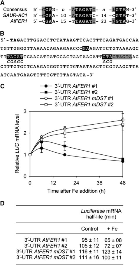
AtFER1 transcript degradation in response to Fe requires the DST cis-acting element present in its 3′-UTR. (A) Sequence alignment of DST sequence of AtSAUR-AC1 (At4g38850) with the DST found in the 3′-UTR of AtFER1. Numbers refers to the numbers of nucleotides between the indicated sequences. Nucleotides in black correspond to those found in the consensus of DST sequences (Sullivan and Green, 1996). (B) Sequence of the 3′-UTR of AtFER1 mRNA. Nucleotides in bold correspond to the stop codon. Nucleotides potentially belonging to a DST element are shown in black. Nucleotides in italic correspond to the mutations introduced in the 3′-UTR AtFER1 mDST constructs. (C) LUC mRNA abundance after Fe addition to transgenic plants. LUC is under the control of the strong 35S-CaMV promoter. The 3′-UTR of AtFER1 containing (3′-UTR AtFER1mDST) or not (3′-UTR AtFER1) mutations in the putative DST motif was introduced downstream of LUC. Two representatives from five independent transgenic lines are presented. For comparing transgenic lines, LUC mRNA abundance was fixed to one before Fe addition. Values and standard errors were obtained from three independent experiments. (D) LUC mRNA half-life in the transgenic lines analysed. Before Fe addition (control), or 3 h after 300 μM Fe-citrate addition (+Fe), cordycepin was added. LUC mRNA half-life was determined as described in the legend of Figure 3. Values and standard errors were obtained from three independent experiments.

