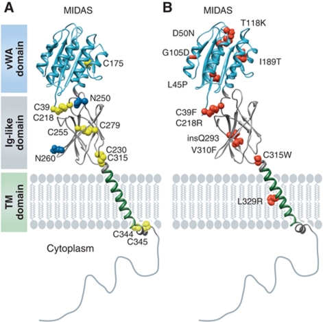
Figure 1.
CMG2 structure and HFS mutations. Schematic representation of the CMG2 structure. The vWA domain corresponds to the crystal structure (1tzn) and the Ig-like domain to our recently published model (Deuquet et al, 2011). (A) Residues in blue represent the N-glycosylation sites and cysteine residues are shown in yellow. (B) The position and identity of reported extracellular missense HFS mutations are mapped onto the CMG2 model. The figure was generated using UCSF Chimera© software (Pettersen et al, 2004).

