Figure 5.
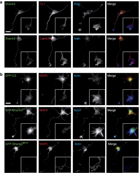
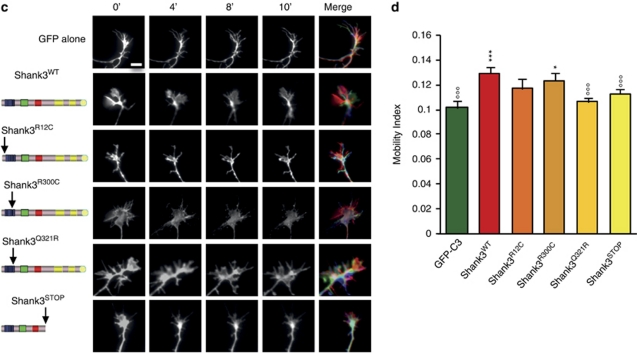
Effects of Shank3WT and mutants on growth cone dynamics. (a) Shank3 is found in the growth cone of neonatal rat-cultured hippocampal neurons on DIV2. Neurons are labeled for Shank3 (green), F-actin (phalloidin; blue) and either tubulin (anti-Tuj1; red) or cortactin (anti-cortactin, red). Scale bars represent 20 μm. Insets represent high magnifications of the axonal growth cone. Shank3 colocalized with F-actin (blue) and with cortactin (red) in axonal growth cones. (b) Localization of Shank3 overexpressed in young neurons. Green fluorescent protein (GFP)–Shank3WT or Shank3STOP or GFP–C3 (green) was transfected in young neurons (DIV0). Immunostaining for MAP2 (red) and actin (phalloidin; blue) revealed that Shank3WT accumulates in growth cones, whereas Shank3STOP is restricted to the cell body. (c) Time-lapse video microscopy of growth cones from rat hippocampal neurons co-transfected with the control (GFP–C3) and Shank3WT or mutated forms. Merge represents three frames with different colors (0 min in green, 5 min in red and 10 min in blue). Scale bars represent 5 μm. (d) Quantification of growth cone motility with the motility index.28 ***P<0.001 and *P<0.05 compared with the control; °°°P<0.001 compared with Shank3WT.


