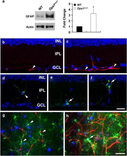
Figure 2.
OPA1 mutation activates both astroglial and microglial cells in Opa1enu/+ mice. (a) Opa1enu/+ mice significantly increased GFAP expression compared with wild-type control mice. Values are mean±S.D. (n=4 retinas/group). *Significant at P<0.05 compared with wild-type control mice. (b and c) GFAP immunohistochemistry. Compared with wild-type control mouse (b), Opa1enu/+ mouse showed increased GFAP immunoreactivity in the GCL (arrowheads, c). (d–f) Iba1 immunohistochemistry. (d) Wild-type control mouse showed Iba1-positive ramified, quiescent microglial cell in the IPL (arrowhead). In contrast, Opa1enu/+ mice showed Iba1-positive microglial cells that have shortened and thickened processes in the IPL (arrow, e) and GCL (arrow, f). (g and h) GFAP (red), Iba1 (green), and Fluorogold (blue) triple labeling. Note that FluoroGold-labeled RGC was engulfed by activated microglia cell in the GCL of Opa1enu/+ mouse (arrow, h). INL, inner nuclear layer; GCL, ganglion cell layer. Scale bars, 20 μm (b–h)

