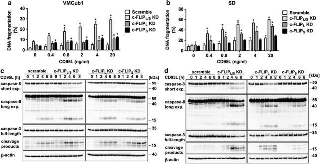
Figure 4.
Knockdown of c-FLIPL and c-FLIPS sensitises urothelial carcinoma cells to CD95L-induced apoptosis. (a and b) For analysis of apoptosis sensitivity, VMCub1 (a) or SD cells (b) stably transduced with the indicated shRNAs against c-FLIPL, c-FLIPS or both isoforms (c-FLIPL/S) were left untreated or stimulated for 16 h with the indicated concentrations of CD95L. The amount of apoptotic cells was quantified by DNA fragmentation analysed by flow cytometry. Data are shown as the mean of four measurements±S.D. Statistical analyses was performed by two-tailed Mann–Whitney U-tests, The symbol * indicates P< 0.05 with respect to scramble controls. (c and d) VMCub1 (c) and SD (d) cells were left untreated or stimulated with 0.4 ng/ml and 0.8 ng/ml CD95L, respectively, for the times indicated. Cleavage of caspase-8 and caspase-3 was analysed by western blot. Expression of β-actin is presented as the loading control

