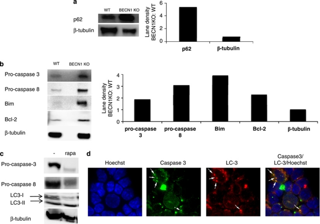
Figure 4.
Beclin 1-deficient CD4+ T cells accumulate cell death-related protein upon activation. WT and 4cre BECN1 fl/fl CD4+ T cells were stimulated for 48 h by anti-CD3 and anti-CD28. At this point, CD4+ cells were re-isolated by positive selection and extract was made for western analysis. The results shown are western blot analyses of p62 (a) and apoptosis proteins, caspase-3, Bim, Bcl-2 and caspase-8 (b). (c) Rapamycin was added at 44 h, and extracts were made at 48 h subjected to western blot analysis. (d) Naïve WT CD4+ T cells were cultured in the Th1 condition for 72 h and then treated with rapamycin for 4 h. Cells were stained with antibodies as indicated. Fluorescence images were obtained. Arrows indicate dots that are positive for both caspase-3 and LC-3

