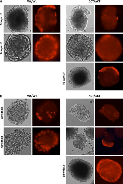
Figure 6.
Ablation of Dido3 delays aggregation-induced primitive endoderm formation. Photomicrographs (gray-tone images) show representative EB obtained by aggregation of Wt/Wt and ΔCT/ΔCT ES cells and subsequent propagation in the absence (w/o) (a) or presence (with) (b) of LIF for the time indicated. Primitive endoderm formation was monitored by immunofluorescence using antibody to the endoderm marker GATA-4 (bright red signal)

