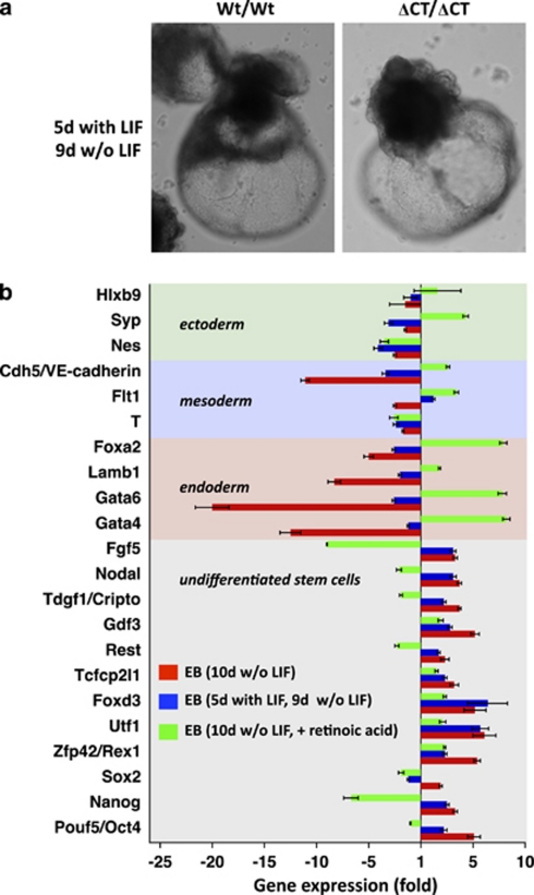
Figure 7.
Impaired differentiation of Dido3 mutant ES cells can be rescued in vitro. (a) Photomicrographs of representative EB obtained by aggregation of Wt/Wt and ΔCT/ΔCT ES cells and subsequent propagation with LIF for 5 days before inducing differentiation by LIF withdrawal for a further 9 days. (b) Quantitative RT-PCR was used to determine the expression of selected markers for undifferentiated ES cells, endoderm, mesoderm and ectoderm in differentiating EB. Differentiation was induced by aggregation of ES into EB and maintenance of EB for a further 10 days in suspension on untreated bacterial plates in medium without LIF (red bars) and supplemented with 0.5 μM retinoic acid (green bars). Alternatively, differentiation was induced by aggregation of ES cells into EB and subsequent maintenance as in a (blue bars). Data show mean values for two experiments±S.E.M. and represent expression of the markers analyzed in ΔCT/ΔCT EB relative to Wt/Wt EB

