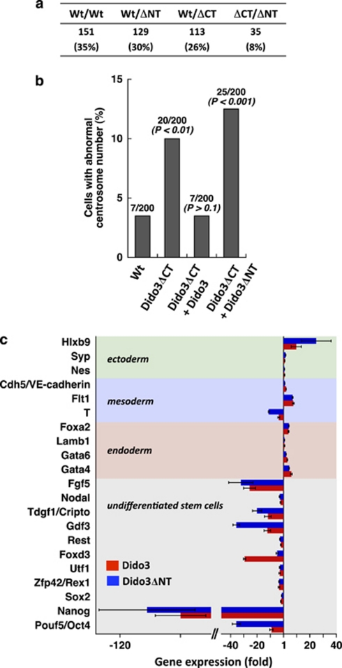
Figure 8.
The role of Dido3 in ES cell differentiation can be distinguished from its function in chromosome segregation. (a) Dido3ΔCT-RFP embryonic lethality was rescued by intercrossing heterozygous Wt/ΔCT with heterozygous Wt/ΔNT mice to obtain a double mutant (ΔCT/ΔNT). Total number of viable pups is shown for each genotype; the contribution of each genotype relative to the total number of mice born is indicated in parentheses. (b) Full-length Dido3 (Dido3) and the N-terminal Dido3 truncation mutant (Dido3ΔNT) were overexpressed in ΔCT/ΔCT ES cells. Centrosome number was determined by staining for γ-tubulin. Cells were scored as positive for centrosome anomalies when more than two or less than one centrosome per cell were detected. The number of cells with centrosome amplification as a percentage of the total number of cells analyzed is shown above each bar. The Fisher exact test was used for statistical comparison with Wt/Wt ES cells. (c) Full-length Dido3 and Dido3ΔNT were overexpressed in ΔCT/ΔCT ES cells. Differentiation was induced by aggregation into EB and maintenance of EB for a further 10 days without LIF. Quantitative RT-PCR was used to determine the expression of selected markers for undifferentiated ES cells, endoderm, mesoderm and ectoderm in ΔCT/ΔCT EB overexpressing full-length Dido3 (red bars) and Dido3ΔNT (blue bars). Data show mean values of two experiments±S.E.M. and represent expression of the markers analyzed in ΔCT/ΔCT EB transfected with Dido3 and Dido3ΔNT, relative to Wt/Wt EB

