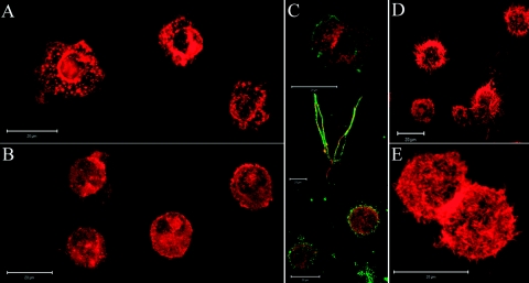
Figure 3.
Cathepsin X enables podosome formation. Immature DCs on Day 5 of differentiation were centrifuged with cytospin (Cytofuge) for 6 min at 1000 rpm onto glass cover slides. Actin was labeled with phalloidin-tetramethylrhodamine B isothiocyanate conjugate (500 ng/ml) for 30 min at room temperature. Podosome formation, present in control, immature DCs (A), is prevented by inhibition of cathepsin X during DC differentiation (B). Adhesion of maturing DCs coincides with β2-integrin activation and colocalization with actin (C). Immature and mature DCs were labeled by centrifugation with cytospin (Cytofuge), whereas maturing, adherent DCs were labeled by seeding immature DCs onto glass coverslips in 24-well plates in the presence of 20 ng/ml LPS and allowing adherence for 20 h. The active form of β2 integrin was labeled with mAb 24 (green fluorescence) and colocalized with actin (red fluorescence) in adherent, mature DCs. Meanwhile, formation of typical dendrites in mature DCs (D) was not inhibited by cathepsin X inhibition (E). Original scale bars represent 20 μm. Fluorescence microscopy was performed using a Carl Zeiss LSM 510 confocal microscope. Images were analyzed using Carl Zeiss LSM image software 3.0.

