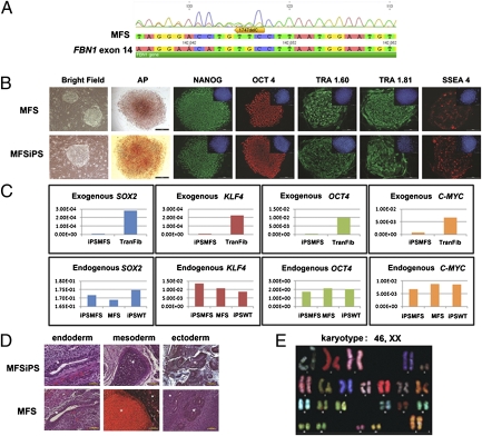
Fig. 1.
Characterization of MFS and MFSiPS cells. (A) DNA sequencing analysis of MFS cells showing a mutation in FBN1 exon 14. (B) Cell morphology of representative MFS and iPS clones by phase contrast (bright field), alkaline phosphatase (AP) staining, and immunofluorescence staining for pluripotent markers: NANOG, OCT-4, TRA-1–60, TRA-1–81, and SSEA-4. (Insets) Nuclear counterstaining performed with DAPI. (Scale bars, 100 μm.) (C) qPCR for the expression of exogenous and endogenous SOX2, KLF4, OCT4, and C-MYC genes. (D) Differentiation of MFS and MFSiPS cells, teratomas containing cells from three germ layers (endoderm, mesoderm, and ectoderm) developed from MFSiPS and MFS cells injected into the dorsal flank of nude mice. Endoderm (gut epithelium), mesoderm, (cartilage), and ectoderm (neuroectoderm) are indicated by white asterisks. (Scale bars, 100–250 μm.) (E) Spectral karyotyping analysis of MFSiPS cells. TransFib, parental transduced fibroblasts.

