Fig. 2.
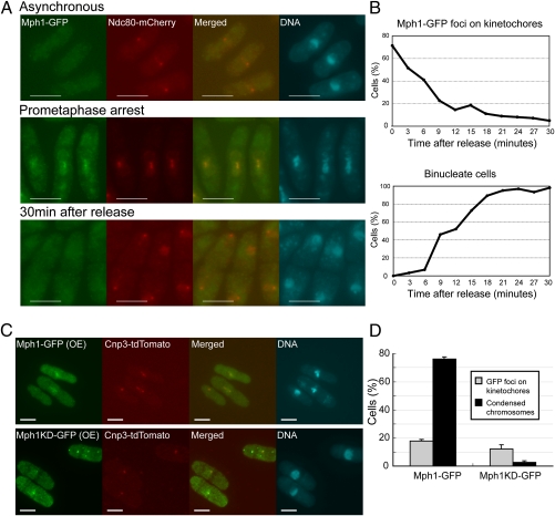
Localization of Mph1. (A) Localization of Mph1-GFP (green) was determined in the cold-sensitive mutant nda3-KM311. The cells were first precultured at 32 °C (asynchronous) and shifted down to the restrictive temperature, 20 °C for 8 h (prometaphase arrest). They were then shifted back to 32 °C to release from the arrest (30 min after release). Ndc80-mCherry (red) was used as a marker of kinetochores. DNA (blue) was visualized by staining with DAPI (4′-6-diamino-2-phenylindole). (Scale bars, 5 μm.) (B) The samples were prepared as in A and the percentages of the cells with Mph1 foci at kinetochores (Upper) and binucleate cells (Lower) were determined after release from the prometaphase arrest. (C) Localization of Mph1-GFP or Mph1-KD-GFP (green) expressed from pREP41 for 18 h at 32 °C was determined with Cnp3-tdTomato (red) as a marker of kinetochores. (Scale bars, 5 μm.) (D) The samples were prepared as in C and the percentages of cells with Mph1 foci on kinetochores (gray bars) and cells with condensed chromosomes (black bars) were determined.

