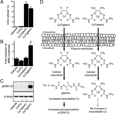
Fig. 1.
Comparative effects of CuCl2, CuII(atsm), and CuII(gtsm). (A) SH-SY5Y cells treated with 10 μM CuCl2, CuII(atsm), or CuII(gtsm) for 1 h before analyzing cells for Cu content by ICP-MS, which does not differentiate between intact CuII(atsm)/CuII(gtsm) and Cu that has dissociated from the respective bis(thiosemicarbazone) scaffolds. (B) Before treating with 500 nM CuCl2, CuII(atsm), or CuII(gtsm) for 6 h, SH-SY5Y cells were transfected with MRE-luciferase construct. Increased bioavailable Cu within the cell upregulates expression of the luciferase reporter, induction of which is measured by luminescence. (C) SH-SY5Y cells treated with 10 μM CuCl2, CuII(atsm), or CuII(gtsm) for 1 h were analyzed for phosphorylated ERK1/2 (pERK1/2). β-Actin levels are shown as a control. (D) Schematic illustrating that although CuII(atsm) and CuII(gtsm) both enter the cell, only CuII(gtsm) is sensitive to the activity of cellular reductants under normal conditions. Increased ERK1/2 phosphorylation is responsive to increases in bioavailable Cu within the cell. Vehicle represents cells treated with the DMSO vehicle used to prepare the Cu compounds. Data are mean values ± SEM, n = 6–12. **P < 0.01 compared to vehicle-treated cells (ANOVA with Tukey’s posttest).

