
Figure 5.
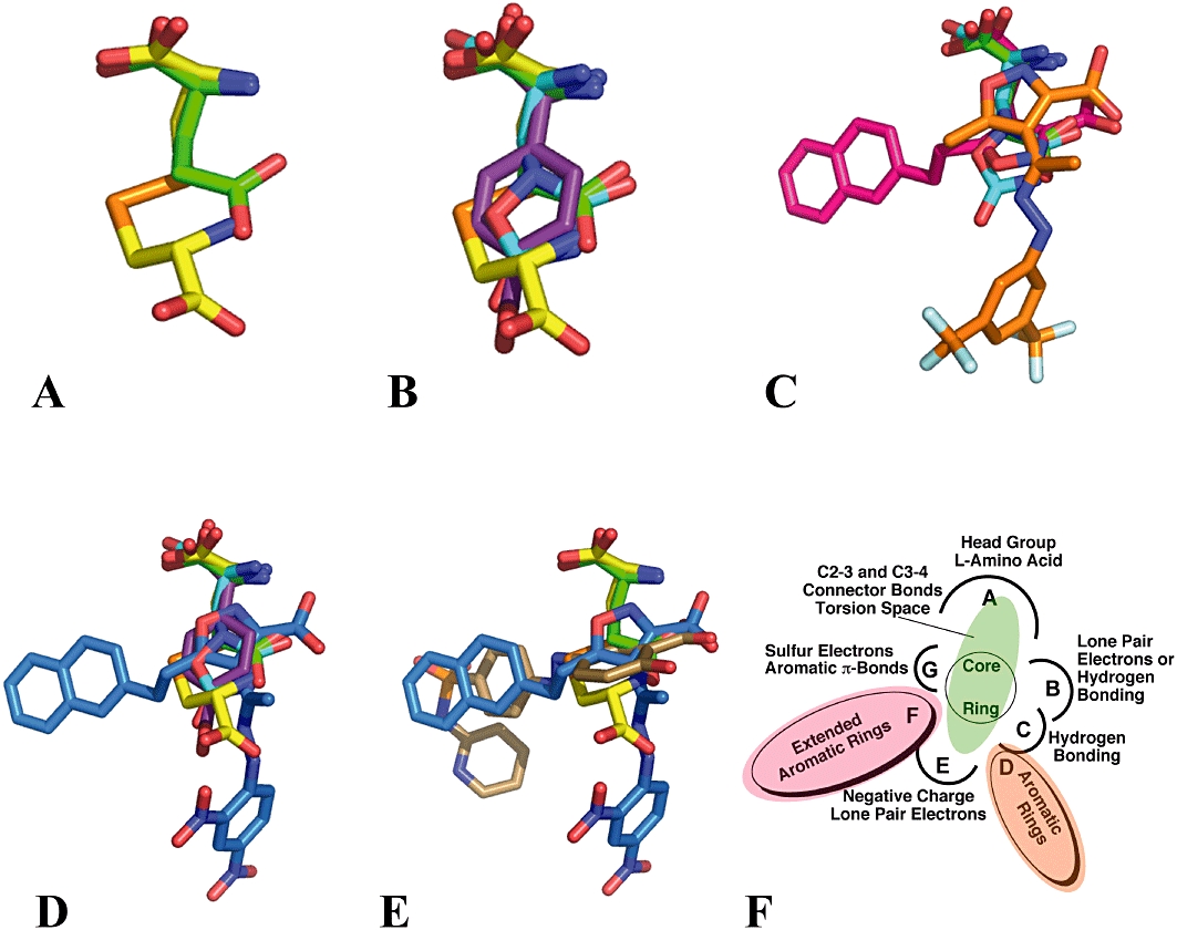
A ligand-based, superposition, 3D pharmacophore model for the substrate binding on Sxc-. A–E. l-Glu in green; l-Cys2 in yellow; QA in teal; 4-S-CPG in purple; NACPA in red; TFMIH in orange; NEIH in blue and SSZ in bronze. F. Pharmacophore binding template.