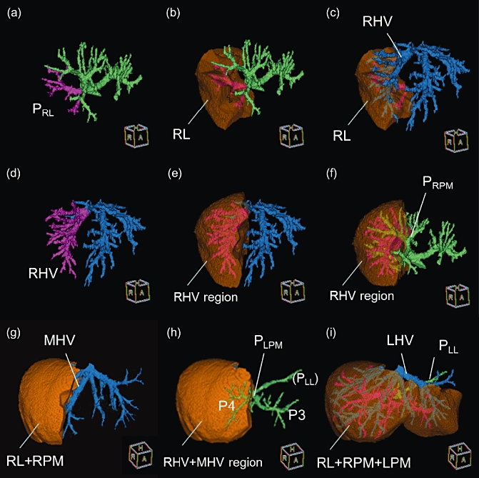
Figure 1.

Step-by-step definitions of the vasculature in livers with right-sided ligamentum teres (RSLT). (a) Three-dimensional configurations of portal branches (green) and the right lateral portal pedicle (PRL) (pink). (b) Visualization of the right lateral sector. (c) When the hepatic veins (blue) are superimposed on the 3D images of right lateral sector, the right hepatic vein (RHV) is visible on the sectoral border. (d) The RHV (pink) is identified. (e) The drainage area of the RHV is then visualized. (f) The right paramedian portal pedicle (PRPM) is visible on the intersectoral plane emerging after visualizing of the RHV. (g) The MHV was confirmed after visualization of the right hemiliver (RL + RPM). RL, right lateral sector; RPM, right paramedian sector. (h) The left paramedian portal pedicle (PLPM) is visible in the umbilical fissure. (i) The left hepatic vein (LHV) was exposed on the sectoral border and the left lateral portal pedicle (PLL) can also be seen behind the LHV
