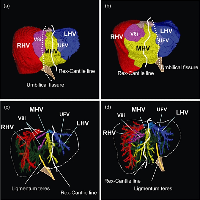
Figure 6.

The distribution of venous drainage areas. (a, c) RSLT case, (b, d) typical liver anatomy. Each venous drainage area is represented in a different colour on the liver surface. The white line represents the simulated Rex-Cantlie line and the dotted line indicates the umbilical fissure. The umbilical fissure is located between the areas of the middle hepatic vein (MHV) and the right hepatic vein (RHV) in the livers with right-sided ligamentum teres (RSLT), while it is usually observed between the MHV and the LHV in the typical liver anatomy
