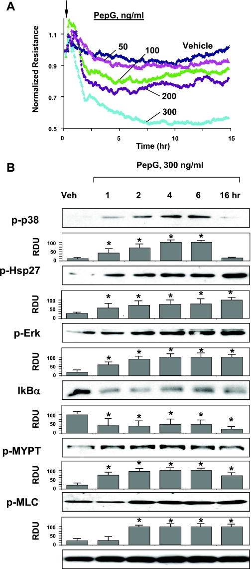
Fig. 2.
Effects of peptidoglycan (PepG) on EC barrier function and inflammatory signaling. A: EC monolayers grown on microelectrodes were treated with PepG (50, 100, 200, or 300 ng/ml, indicated by arrow) and used for permeability measurements. B: HPAEC were stimulated with PepG (300 ng/ml) for indicated periods of time. Phosphorylation of p38, Hsp27, ERK1/2, MYPT, and MLC was determined by Western blot with corresponding phospho-specific antibodies. Degradation of IκBα was detected using pan IκBα antibodies. Equal protein loading was confirmed by determination of β-tubulin content in total cell lysates. Results are representative of three to six independent experiments. Result of densitometry are shown as means ± SD. *P < 0.05, compared with vehicle control.

