
Fig. 7.
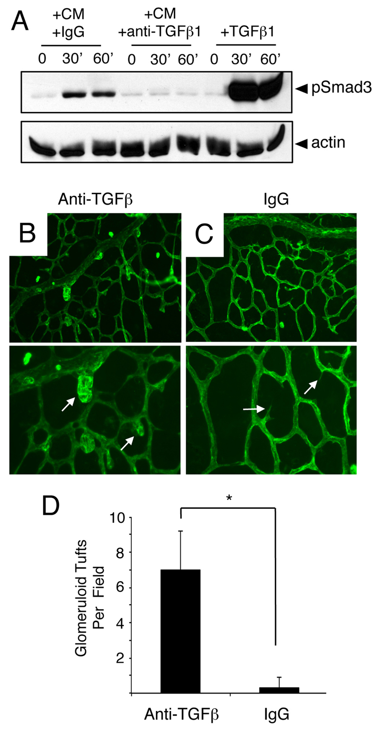
Anti-TGFβ neutralizing antibodies induce acute retinal angiogenesis defects. (A) Primary brain microvascular endothelial cells were treated for varying times (0, 30 or 60 minutes) with 1 ng/ml TGFβ1 (+TGFβ) or retinal astrocyte conditioned media (CM) containing anti-TGFβ neutralizing antibodies (+anti-TGFβ) or control IgGs (+IgGs). Note that treatment of endothelial cell cultures with TGFβ1 or CM+IgG leads to a time-dependent increase in Smad3 phosphorylation. By contrast, anti-TGFβ neutralizing antibodies inhibit Smad3 phosphorylation in endothelial cells. (B,C) P9 mouse pups were anesthetized and injected intraocularly with anti-TGFβ neutralizing antibodies (B) or control IgGs (C). Retinas were fluorescently labeled with IsoB4-Alexa488 to visualize blood vessels. Note the abnormal blood vessel morphologies in P9 mice injected with anti-TGFβ blocking antibodies (arrows in B). Images are shown at 200× magnification in the upper panels and at 400× magnification in lower panels. (D) Quantification of retinal angiogenesis pathologies in mice intraocularly injected with control IgGs or anti-TGFβ antibodies. *P<0.001. Error bars represent s.d.