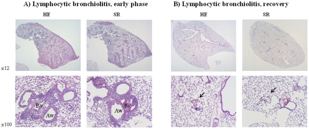
Figure 6. Histology of allografts showing lymphocytic bronchiolitis lesions.
A) early after transplantation (2w) and B) late after transplantation (12w). Left side: HE staining, right side: Sirius Red (SR) staining. 2w after transplantation allografts showed enlargement of broncho-vascular axes with infiltration of lymphocytes around airways (Aw) and blood vessels (Bv). Sirius Red staining shows collagen both perivascular as peribronchial. After 12w, lungs completely recovered with some leftover damage seen in pigmented macrophages (arrow).

