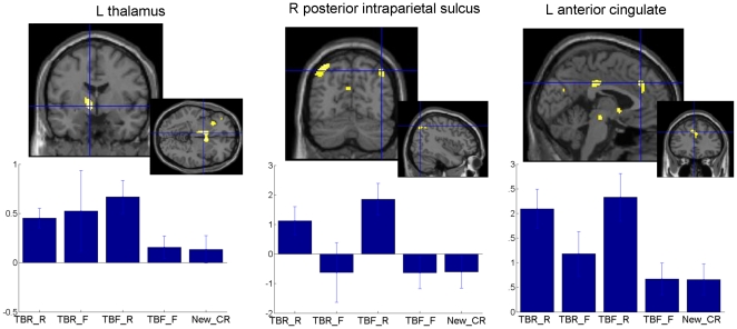
Figure 3. Cerebral areas associated to retrieval of TBF information ( Table 4 ).
Larger brain responses for TBF-R than TBF-F information in the left thalamus (left), right posterior intraparietal sulcus (middle) and left anterior cingulate (right). Functional statistical results (puncorrected<0.001) are overlaid to a canonical structural image. Activity estimates (arbitrary units) are displayed for the different conditions. TBR-R: items associated to a TBR instruction and subsequently recognised; TBR-F: items associated to a TBR instruction and subsequently forgotten; TBF-R: items associated to a TBF instruction and subsequently recognised; TBF-F: items associated to a TBF instruction and subsequently forgotten; New_CR: correct rejection of items not presented during the encoding session.

