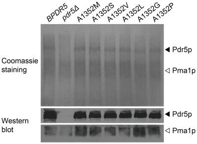
Figure 5. Membrane localization of Pdr5p and its mutant variants.
Isolated plasma membranes (15 µg/lane) were electrophoresed on an SDS–8% polyacrylamide gel and visualized with Coomassie blue (top panel). Pdr5p (middle panel) and Pma1p (bottom panel) were immunodetected with specific antibodies as described in the materials and methods section. Filled arrowheads indicate Pdr5p and open arrowheads indicate Pma1p. Coomassie blue stained SDS-PAGE gel (upper panel) and western blot (lower panel) of the same strains are shown.

