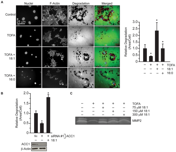
Figure 5. Fatty acid addition rescues gelatin degradation in 3T3-Src cells treated with TOFA.
(A) Gelatin degradation of 3T3-Src cells seeded on glass cover slips coated with AlexaFluor-488 conjugated gelatin in the presence of vehicle, TOFA (30 µM), or TOFA with 18∶1 or 16∶0 fatty acid. Nuclei were visualized by staining with DAPI and actin was visualized with Alexafluor594-phalloidin. (B) Quantification of gelatin degradation by 3T3-Src cells transfected with scrambled (Sc) or siRNA #1 against ACC1 and supplemented with 18∶1 fatty acid for 24 hours. (C) Gelatin zymography of secreted MMP-2 in condition media from 3T3-Src cells treated with vehicle, TOFA (30 µM), or TOFA supplemented with 18∶1 fatty acid for 48 hours. Scale bar, 25 µm. *p≤0.05.

