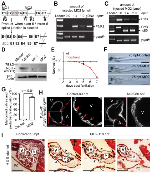
Fig. 2.
Npnt knockdown disrupts heart development and is lethal. (A) Scheme of the effect of npnt splice-inhibitory morpholino MO2. Exons are represented by boxes and introns by lines. Dotted lines indicate the region targeted by MO1 or MO2 and arrows indicate RT-PCR primer positions. Exon E3 could not be detected. (B,C) RT-PCR analysis of mRNAs from control and MO2-injected zebrafish embryos at 52 hpf using npnt primers as indicated in A and gapdh primers (loading control) (n=3). MO2 inhibited npnt pre-mRNA splicing, resulting in intron insertion (B) and exon E5 deletion (C). (D) Western blot analysis of control and MO1- or MO2-injected embryos at 52 hpf (n=3). Both morpholinos efficiently knocked down the Npnt protein level. (E) Npnt knockdown resulted in 89±7.9% (mean ± s.e.m.) lethality at 7 dpf. (F) Lateral view of control, MO1- or MO2-injected embryos at 75 hpf. MO1 or MO2 injection resulted in pericardial edema (arrowheads). (G) Quantitative analysis. Valve formation and trabeculation are perturbed in 86±3.9% (mean ± s.e.m.) of morphant hearts at 110 hpf. (H) Confocal images of hearts from control and MO2-injected embryos from transgenic Tg(myl7:EGFP-HsHRAS)s883 zebrafish at 80 hpf suggesting that the initiation of trabeculation is perturbed in npnt morphants. Red arrows indicate trabeculae. (I) Hematoxylin and Eosin (H&E)-stained sagittal sections of hearts from control and MO2-injected embryos at 110 hpf. In contrast to morphants, control embryos develop proper atrioventricular (AV) valve leaflets (white oval). Black arrowheads indicate the AV boundary; green arrows indicate expanded cardiac jelly. A, atrium; V, ventricle; PC, pericardium. Scale bars: 500 μm in F; 50 μm in H,I.

