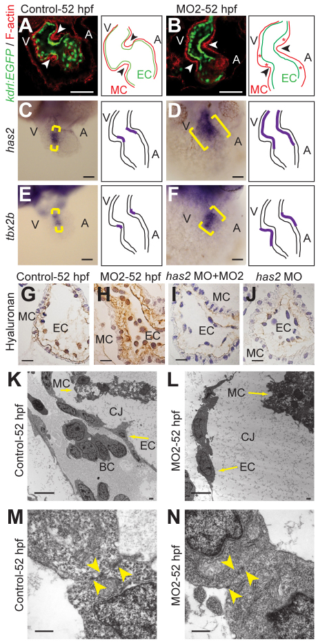
Fig. 5.
Npnt knockdown causes cardiac jelly expansion and expanded expression of has2 and tbx2b. (A,B) Confocal sections from whole-mount F-actin-stained (red) control (A) and MO2-injected (B) Tg(kdrl:EGFP)s843 zebrafish embryos at 52 hpf. The space between endocardium and myocardium was increased at the AV boundary (arrowheads) and throughout the chambers (red asterisks) in npnt morphants. (C-F) Sections of 52 hpf control and MO2-injected embryos after whole-mount in situ hybridization for has2 and tbx2b expression (brackets). Note that the expression of these genes is expanded in npnt morphants. (G-J) Sections (4 μm) of control, MO2-, has2 MO-, or has2 MO + MO2-injected zebrafish embryos at 52 hpf stained for hyaluronan (brown) and counterstained with Hematoxylin (blue). (K-N) Transmission electron microscopy of hearts (K,L) and endocardial cells (M,N) from control and MO2-injected embryos at 52 hpf. Arrowheads indicate junctional complexes. A, atrium; BC, blood cell; CJ, cardiac jelly; EC, endocardium; MC, myocardium; V, ventricle. Scale bars: 50 μm in A-F; 10 μm in G-J; 5 μm in K,L; 1 μm in M,N.

