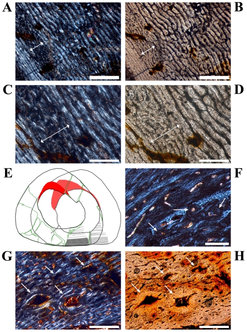
Figure 6. The Mark of Initial Sexual Maturity (MISM) as well as interior details of the anterior corner in large sampled femora of Dysalotosaurus.
A–D: SMNS F1, A – Part of the posterior bone wall, under polarized light, with the most external fast growing zone (double-headed arrow) and the transition to the thick, non-cyclical slow growing area externally (centre and right of the image). This transition is the MISM. B – The same as in A under normal light. C – Magnification of the top left of A under polarized light. The MISM is again at the right end of the double-headed arrow. Note that the MISM is not a sharp line but just another transition from fast to slower growth without any further fast growing zones towards the periphery. D – The same as in C under normal light. E: GPIT/RE/3414 (in front) and GPIT/RE/3588 (in the back), the sketches demonstrate the perfect overlap of the zonation as well as the MISM in both large femora. The slow growing zones are shaded in the back and their external rim is marked in the front. The dashed lines within the thick external slow growing zone (shaded in both representing growth after reaching sexual maturity) mark unsecured growth cycles. F: GZG.V 6590 28, Close up of the border between the CCCB wedge internally (bottom right) and the primary bone tissue externally within the anterior corner. Secondary osteons are marked by arrows. Note the knitted pattern of the primary bone tissue. G–H: GZG.V 6211 22, G – Internal part of the anterior corner close to the CCCB wedge (starts at the lower right) with knitted pattern of the bone tissue internally and some scattered secondary osteons (arrows) still under development, under polarized light. H – The same as in G under normal light. Scale bars = 1 mm in A–B. Scale bars = 500 µm in C–D, G–H. Scale bars = 200 µm in F.

