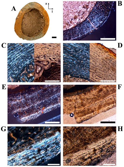
Figure 8. Bone histology and growth cycles in juvenile femora of Dysalotosaurus.
A–D: GZG.V 6379, A – Orientated overview (a = anterior, l = lateral) under normal light. Note the wide marrow cavity compared to the bone wall thickness in this early juvenile specimen. B – Magnification of the posterolateral corner under polarized light with only a weak indication of the Posterolateral Plug. The knitted pattern of the bone tissue with mainly longitudinal primary osteons is dominant. C – Magnification of the interior of the anterior corner medially, under both polarized and normal light, with a CCCB wedge under development and the typical knitted pattern of the primary bone tissue. D - Magnification of the interior of the anterior corner laterally, under both polarized and normal light, with the typical knitted pattern of the primary bone tissue. The vascular pattern changes already to more circumferential primary osteons towards the periphery. E–H: GZG.V 6590, E – Three slow growing zones are well visible under polarized light. The Posterolateral Plug starts at the left edge of the image. F – The same as E under normal light. G – Magnification of the utmost slow growing zone with an annulus at its interior border. H – The same as in G under normal light. Scale bars = 1 mm in A–B, E–F. Scale bars = 500 µm in C–D. Scale bars = 200 µm in G–H.

