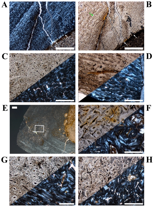
Figure 9. Vascular patterns and tissue types in a Dysalotosaurus tibia.
A–H: Large tibia SMNS T3, A – Internal part of the lateral bone wall with laminar to sub-plexiform bone tissue under polarized light. Transversely oriented bone fibers dominate. The knitted pattern is visible at the right close to the marrow cavity. A thick endosteal layer is marked by white arrows. B – The same as in A under normal light. The external border of the prominent slow growing zone of A is also well visible here (green arrow). C – Strongly unordered primary osteons in a weakly birefringent woven matrix within the medioposterior corner under both polarized and normal light. D – Well organized primary osteons in a strongly birefringent almost parallel-fibered matrix at the outer edge of the lateral wall under both polarized and normal light. E – Overview of the anterolateral corner (here anterior to the bottom and lateral to the left) under polarized light. Note the whirl-like Anterolateral Plug within this corner, which interrupts the usual bone tissue, and the wedge of CCCB to the right at the marrow cavity. F – Partial close up of the CCCB wedge with the usual continuous lamellar bone and some interrupting secondary osteons (arrows), under both polarized and normal light. G – Close up of the border between CCCB (upper right) and primary bone tissue (lower left), under both polarized and normal light. The latter strongly resembles the juvenile knitted pattern. H – Magnification of the framed part in E showing an area within the Anterolateral Plug, under both polarized and normal light. Secondary osteons are marked with arrows. Scale bars = 1 mm in A–B, E. Scale bars = 500 µm in C–D, F, H. Scale bars = 200 µm in G.

