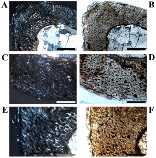
Figure 12. Bone histology of the smallest preserved tibia of Dysalotosaurus.
A–F: Early juvenile tibia GPIT/RE/3795, A – Overview of the anterolateral corner under polarized light. CCCB and the Anterolateral Plug are absent. The interior part of that corner is altered by preservation (see also Fig. S1). B – The same as in A under normal light. C – The posterior wall is well vascularized and the primary osteons are plexiform to reticular in arrangement. The degree of organization as well as of the birefringence seems to increase towards the external surface, under polarized light. D – The same as in C under normal light. E – Magnification of A under polarized light. F – Magnification of B under normal light showing many simple vascular canals oriented radially. Scale bars = 1 mm in A–B. Scale bars = 500 µm in C–F.

