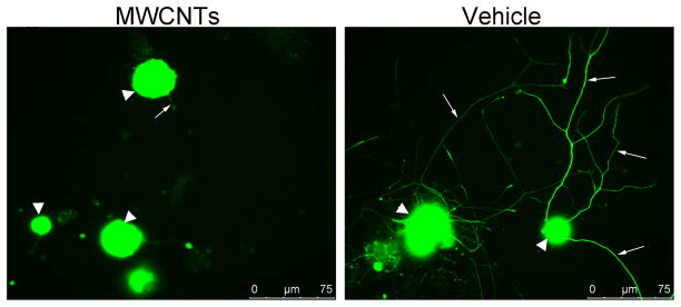
Figure 1. Indirect immunofluorescence of DRG neurons 24 hours after plating.
Indirect immunofluorescence was performed against neuronal β-tubulin using primary mouse anti-TUJ1 antibody, and anti-mouse FITC secondary antibodies. Left panel shows DRG neurons incubated overnight with 10ug/ml of MWCNTs in 10% of surfactant in saline. Right panel shows control DRG neurons incubated with vehicle (10% of surfactant in saline). Big white arrowheads point at DRG neurons. Small arrows point at axons.

