
Figure 1.
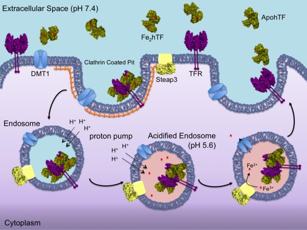
Endocytic hTF/TFR cycle. Iron bound hTF (green) in the blood binds to the specific TFR (purple) with nM affinity at the cell surface (pH 7.4). The hTF/TFR complex is endocytosed in a clathrin-coated pit. Within the endosome, the pH is lowered to ~5.6 causing iron to be released from hTF to an, as yet, unidentified chelator. Fe3+ is reduced to Fe2+ by the ferrireductase Steap3 (yellow) within the endosome. The Fe2+ can then be transported out of the endosome via the divalent metal transporter DMT1 (blue) for use throughout the cell. The apohTF remains tightly bound to the TFR at pH 5.6 and is recycled back to the cell surface. Upon exposure to the slightly basic pH (7.4), apohTF is released or displaced from the TFR and free to bind more Fe3+.