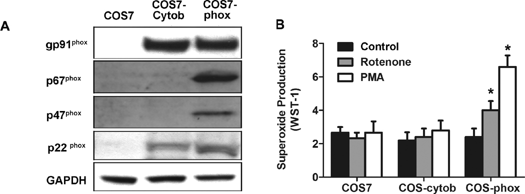
Fig. 3. Both membrane and cytosolic subunits were required for rotenone-induced extracellular superoxide release.
(A) Western blotting shows expression of PHOX subunits in COS7, COS-cytob558 (COS7 cells stably transfected with flavocytochrome b558), and COS-phox cells (COS7 cells stably transfected with both flavocytochrome b558 and cytosolic subunits p47phox and p67phox). (B) Extracellular superoxide release was measured by the SOD-inhibitable reduction of WST-1 after treatment with vehicle, rotenone (10 nM), or PMA (40 nM). Data were shown as mean ± SEM from three independent experiments in triplicate. *, P < 0.01 compared with corresponding vehicle-treated controls.

