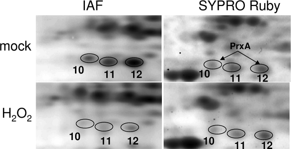
Figure 6.
Comparison of the IAF-labeled protein spots and SYPRO Ruby-stained protein spots from a portion of 2-D gels from the blocking-IAF labeling method. The numbered protein spots correspond to those in Figure 3B. Note the reduced IAF labeling of Spots 10, 11, and 12 in the H2O2-treated sample compared to that in the mock-treated sample. Spot 10 and Spot 12 were identified as PrxA. Staining of the gels with SYPRO Ruby revealed that the abundance of these proteins was not changed by the oxidant treatment.

