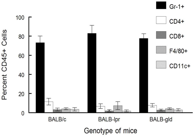
Figure 5.
Inflammatory infiltrate from BALB/c, BALB-lpr, and BALB-gld mice does not indicate strain-specific differences. HSV-infected corneas were removed at days 17 and 23 from mice with severe HSK disease and disaggregated into single-cell suspensions and stained with anti-CD45, CD4, CD8α, Gr-1, CD11b, CD11c, and F4/80 mAb. Cells were analyzed by flow cytometry. Data represents 4 to 6 corneas per group. No significant differences were seen for any particular determination between strains of mice tested.

