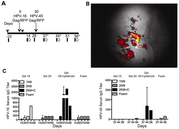
FIGURE 1.

HPV transduces macaque epithelium and induces antibodies to HPV. Cynomolgus macaques were vaccinated intra-vaginally with HPV pseudovirions expressing either RFP or SIV Gag. (A) Schematic showing the design of the study, eight cynomolgus macaques were vaccinated with HPV16 and HPV45, thirty days apart. Blood (black bars) and tissues (diamonds) were sampled pre and post vaccination. In order to obtain sufficient samples to analyze the female genital tract, animals were serially sacrificed at 37, 44, or fifty six days post vaccination (stars). (B) Forty-eight hours post vaccination a camera fitted to an endoscope was used to visualize the cervix and vaginal tract. Successful HPV vaccination was confirmed by the detection of RFP in each animal. Shown is the level of in vivo fluorescence from HPV-RFP PsVs measured in a representative animal. (C) Serum IgG to the HPV16 and 45 capsid L1 measured fourteen-fifty six days post vaccination. The vaginal epithelia were disrupted by 4 methods using nonoxynol 9 (N9). N9 was prepared as a 10% gel and delivered once (Gel 1X white bars), twice, (Gel 2X hatched bars), or twice in combination with abrasion using a cytobrush (Gel 2X+cytobrush, black bars). In addition, we used a 12.5% foam application of N9 also given twice (Foam striped bars).
