FIGURE 4.

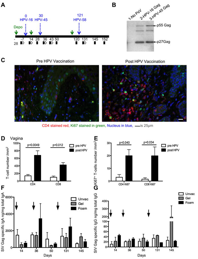
HPV PsVs are immunogenic in rhesus macaques, recruit CD4+ and CD8+ T-cells to the site of vaccination and induce vaginal humoral responses. (A) Schematic showing the vaccination and sampling schedule in rhesus macaques. Macaques were vaccinated on days 0, 30, and 121 with HPV16, 45, and 58, respectively. Blood (black squares) and tissues (white diamonds) were collected pre and post HPV vaccination. (B) Western blot showing the expression of SIV Gag polyprotein p55 and processed p27 protein in 293TT-cells transduced with HPV16 and HPV45 Gag-Pro constructs (lanes 2 & 3). Non-transduced 293TT lysates was used as a negative control (lane 1).
(C) Vaginal biopsies were obtained prior to and one-week post HPV vaccination, and paraffin embedded. A representative example of immunohistochemical staining performed on embedded tissue, stained for CD4 (red) Ki67 (green) and nuclear material stained by dapi (blue) prior to (left) and post (right) HPV vaccination. (D) The absolute number of CD4+ and CD8+ T-cells enumerated from vaginal biopsies pre (white bars) and post vaccination (black bars). A repeated measure analysis of variance demonstrated that the difference is statistically significant (p=0.0049 and p=0.012). (E) The absolute number of Ki67+ CD4+ and Ki67+ CD8+ cells in vaginal biopsies pre and post HPV vaccination. A repeated measure analysis of variance demonstrated that the difference is statistically significant (p=0.040 and p=0.034) (F) SIV Gag-specific IgA as a function of total IgA in vaginal secretions. (G) SIV Gag-specific IgG as a function of total IgG in vaginal secretions.
