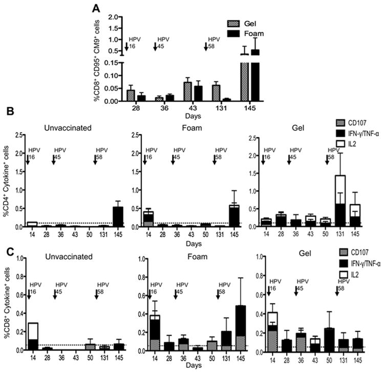
FIGURE 5.

SIV specific Memory T-cell responses to HPV PsVs (A) Antigen experienced CD95+CD28 (+/-) CD8+ T- cells from PBMC’s were gated and the frequency of SIVGagCM9+ cells assessed post HPV vaccination. Animals treated with N9 foam are depicted in hatched bars, while N9 gel treated animals are in black bars. Arrows above indicate the times when each HPV vaccine was administered. (B) Intracellular cytokine staining indicating the frequency of CD107+(hatched), IL-2 (white), IFN-γ and/or TNF-α (black) producing CD4+ T-cells from Gag stimulated PBMC’s in unvaccinated (left) foam treated (middle) and gel treated (right) animals. (C) Intracellular cytokine staining indicating the frequency of CD107+, IL-2+, IFN-γ and/or TNF-α producing CD8+ T-cells from Gag stimulated PBMC’s.
