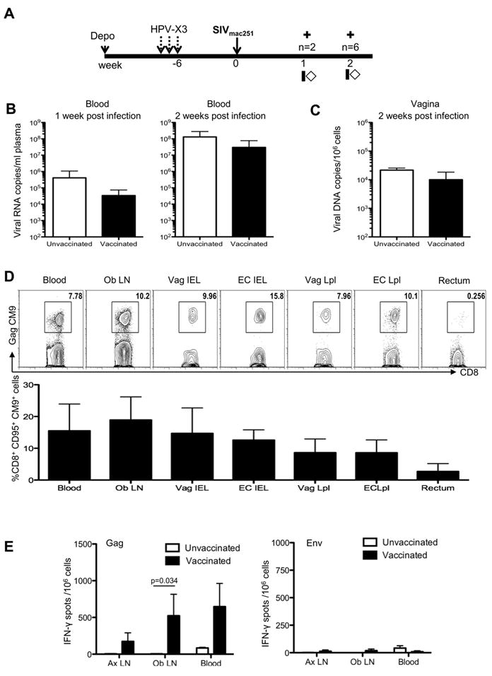
FIGURE 6.

Intravaginal exposure to SIV induces an expansion of vaccine-primed immune responses and does not exacerbate virus replication. (A) Study design showing SIV exposure six weeks post the final HPV vaccine. Animals were sacrificed at one or two weeks post infection (indicated by crosses), and blood and tissues collected at sacrifice. (B) Plasma viral load in unvaccinated and vaccinated macaques one and two weeks post infection. (C) Cell associated viral load in the vagina quantified as the number of SIV DNA copies/106 cells. (D) Representative flow cytometric plots (top panel) showing the frequency of SIVGagCM9 positive memory CD8+ T-cells in the blood, genital draining obturator lymph node, cervix, vagina, and rectum two weeks post challenge. Both intraepithelial and lamina propria CD8+T-cells were obtained from the female genital tract. The bottom panel shows the average frequency of SIV specific Gag CM9 CD95+CD8+ T-cells from vaccinated animals. (E) Average IFN-γ producing spot forming mononuclear cells from axillary, obturator lymph nodes, and blood after stimulation with overlapping peptides spanning SIV Gag (left) or SIV Env (right) from vaccinated and unvaccinated controls, two weeks post challenge. A repeated measure analysis of variance demonstrated that the difference in IFN- γ production from the obturator lymph node between vaccinated and non-vaccinated animals is statistically significant (p=0.034).
