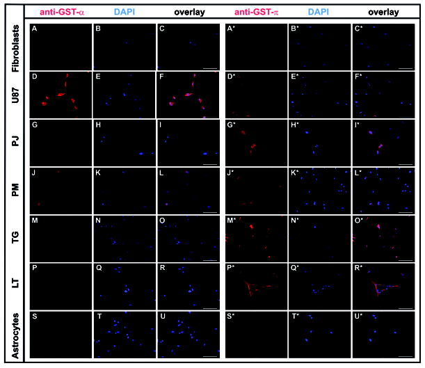
Figure 4.

Expression of GST-α and -π in U87 glioma cells, four selected primary glioblastoma cell lines (PJ, PM, TG, LT), and in fibroblasts and astrocytes was assessed by immunocytochemistry. Cells were fixed, permeabilized, and incubated with specific primary antibodies and fluorescence labelled secondary antibodies for imaging as detailed in Materials and Methods. Photomicrographs taken on a Zeiss Axio Observer Microscope, 20X magnification, scale bar represents 100 μm. Photomicrographs in columns 1 and 4 show immunostaining for GST- α and -π, respectively, columns 2 and 5 show nuclear staining with DAPI, columns 3 and 6 composites with overlay of both images.
