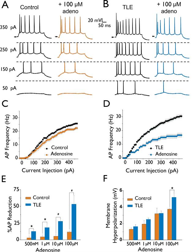
Figure 1. mEC layer II stellate neurons In TLE are hyperexcltable and more sensitive to Inhibition by adenosine than control neurons.
A and B: Membrane properties and neuronal excitability were recorded from both control (A) and TLE (B) mEC layer II stellate neurons (left traces). APs were evoked using current injection steps from -20 to 470 pA. Shown are the responses to 50, 150, 250, and 350 pA current injections. Membrane properties and excitability were again recorded in control (A) and TLE (B) neurons following a 5 minute bath application of 100 |jM adenosine (right traces). C and D: Current-frequency plots showing reduction of AP frequency by adenosine (100 μM) in control neurons (C) and TLE neurons (D). E: Bar chart representing a concentration dependent inhibition of AP frequency measured at a depolarizing current injection step of 470 pA for both control and TLE neurons. Note the profound inhibition of AP frequency in TLE at all doses tested. F: Bar chart demonstrating the average dose-dependent effect of adenosine application on membrane hyperpolarization. Data points represent means ± S.E.M. * P<0.05, # P<0.005.

