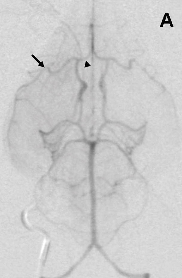
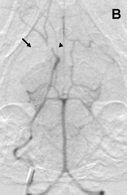
Figure 1.


Rabbit Angiography. Subselective magnification angiograms of the internal carotid artery demonstrate (A) the Circle of Willis and the middle cerebral artery (MCA) and anterior cerebral artery (ACA) (arrow and arrowhead, respectively) and (B) occlusion of the MCA and ACA following the injection of three embolic spheres.
