
Figure 4.
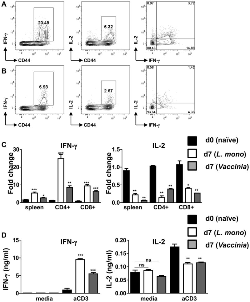
L. monocytogenes- and vaccinia virus-specific CD4+ T cells produce limited amounts of IL-2.
WT mice (five animals per group) were infected with (A) L. monocytogenes (104 CFU per mouse) or (B) vaccinia virus (106 PFU per mouse), and the ability of CD4+ T cells to secrete IFN-γ and IL-2 was analyzed by flow cytometry after restimulation with 0.5 ug/ml αCD3 for 5 hr in the presence of GolgiPlug. (C) Expression levels of IFN-γ (left panel) and IL-2 (right panel) were analyzed by real-time PCR in naïve (black bars), L. monocytogenes (open bars) and vaccinia virus (grey bars) infected mice on day 7 post infection. (D) IFN-γ and IL-2 were measured by ELISA in unstimulated (media) or 0.01 ug/ml αCD3 restimulated splenocytes isolated from naïve (black bars), L. monocytogenes (open bars) and vaccinia virus (grey bars) infected mice. The data shown are representative of four independent experiments. * P< 0.05; ** P< 0.01, *** P< 0.001.